

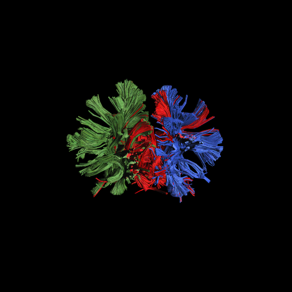
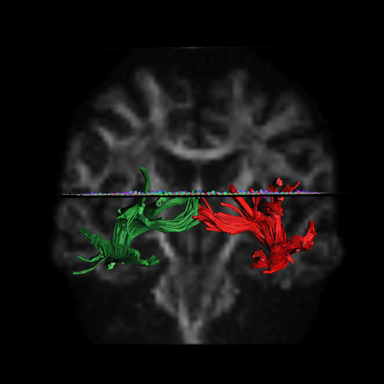
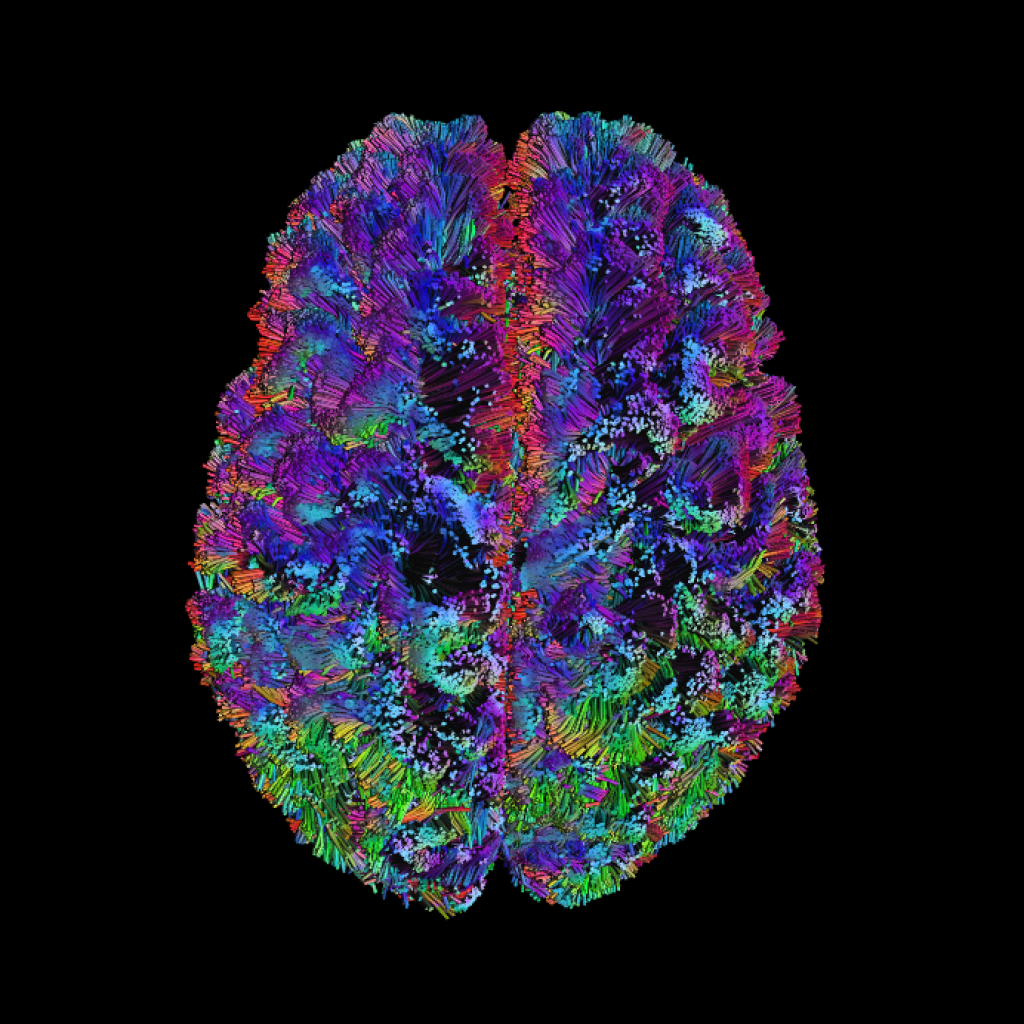
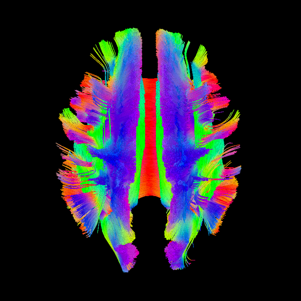
The CfNN Neuroimaging Core supports projects across all Focus Areas. It provides our VA scientists with access to advanced software and hardware for the acquisitions of higher resolution MR images and detection of weaker signals, enabling clearer boundary detection between brain areas and different tissues (e.g., gray-white matter) and more sensitively revealing activation during functional MRI. These enhancements supported through CfNN have also improved the quality of clinical imaging at the VA Providence Healthcare System (VAPHS). Our Core facilitates access for our VAPHS Investigators to the Brown Magnetic Resonance Facility* and its MRI system, which increases scan availability and capability, as it includes some additional advanced technical features not available at VAPHS for research.
Please visit our VA SharePoint** CfNN Core A – Home (sharepoint.com) for more information, or contact Hannah Swearingen at [email protected]
**Sharepoint site only accessible on VA network
Affiliate Laboratories
Barredo Lab – Brown Clinical Neuroimaging Research Core


Computational Neuroscience